Papp.Schneider1993
P. P. Papp
D. K. Chattoraj, and T. D. Schneider.
Information analysis of sequences that bind the replication initiator
RepA.
J. Mol. Biol.
233:219-230, 1993.
https://doi.org/10.1006/jmbi.1993.1501
https://alum.mit.edu/www/toms/papers/helixrepa/
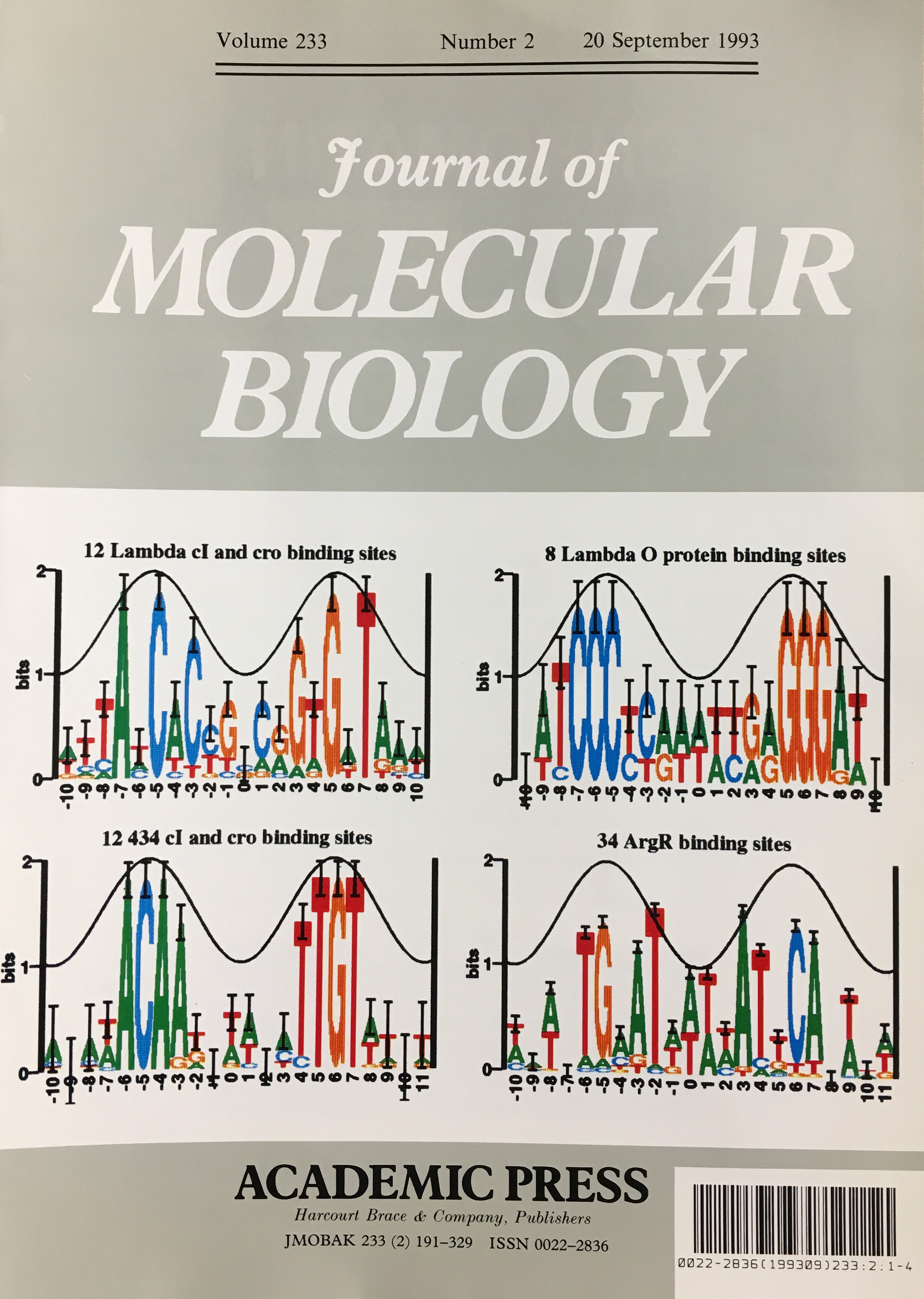
The tall letters represent the highly conserved bases in DNA binding sites of several prokaryotic repressors and activators. Conservation is strongest where major grooves of the double helical DNA (represented by crests of a cosine wave) face the protein. This shows that conservation analysis alone can be used to predict the face of DNA that contacts the proteins.

Schneider-walker1997
T. D. Schneider.
Sequence walkers: a graphical method to display how binding proteins
interact with DNA or RNA sequences.
Nucleic Acids Res.
25:4408-4415, 1997.
https://doi.org/10.1093/nar/25.21.4408
https://alum.mit.edu/www/toms/papers/walker/
erratum: NAR 26(4): 1135,
1998.
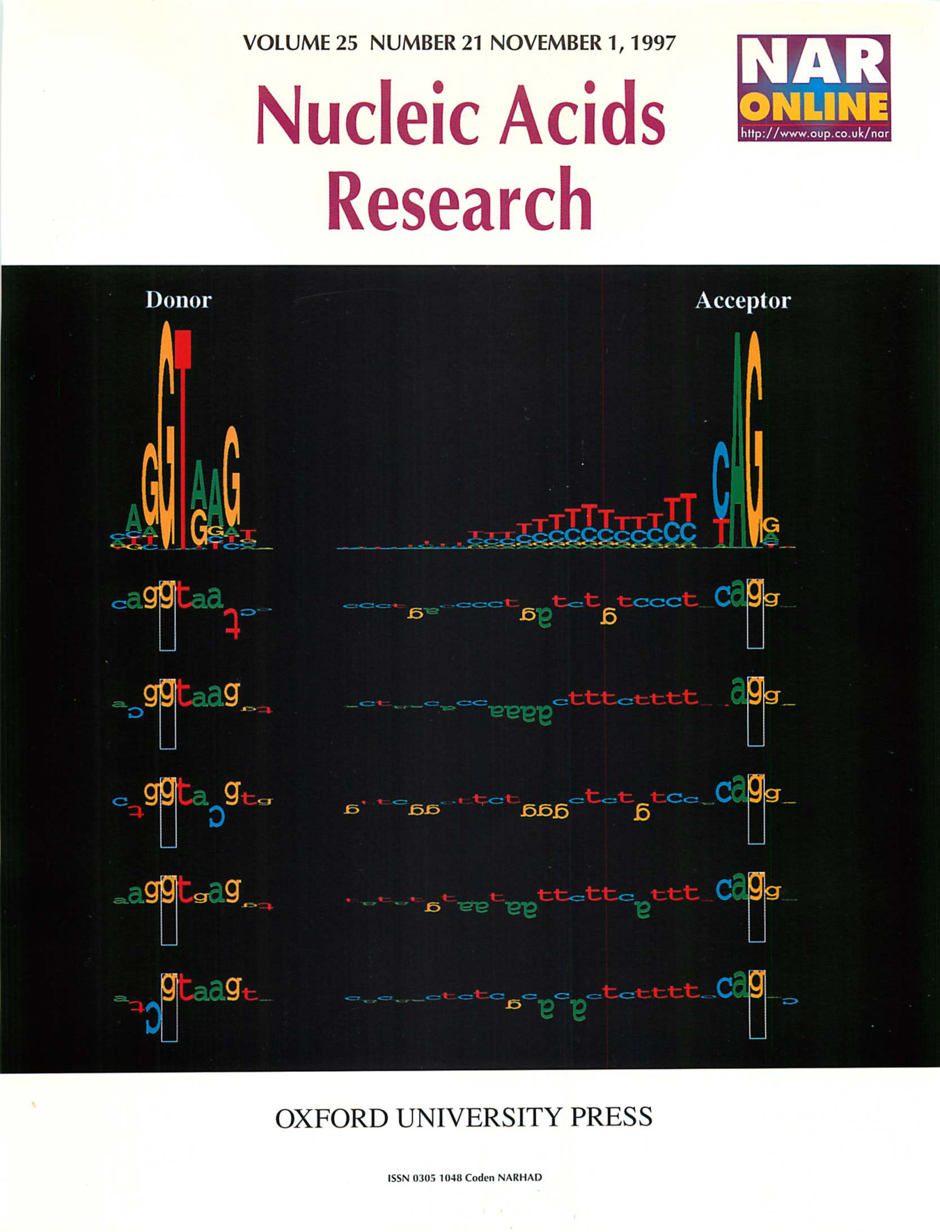
Sequence logos and walkers. At the top of the figure sequence logos for human donor and acceptor sites depict the DNA sequence conservation by stacks of letters. Within each stack, the height of each letter is proportional to the base frequency at that position in the binding site. The height of each stack is the sequence conservation in bits of information. Below the sequence logos five examples of individual binding sites are shown as walkers. The height of each letter shows its contribution to the average sequence conservation in the sequence logo on the top.

Schneider.baseflip.2001
T. D. Schneider.
Strong minor groove base conservation in sequence logos implies DNA
distortion or base flipping during replication and transcription initiation.
Nucleic Acids Res.
29:4881-4891, 2001.
https://doi.org/10.1093/nar/29.23.4881
https://alum.mit.edu/www/toms/papers/baseflip/
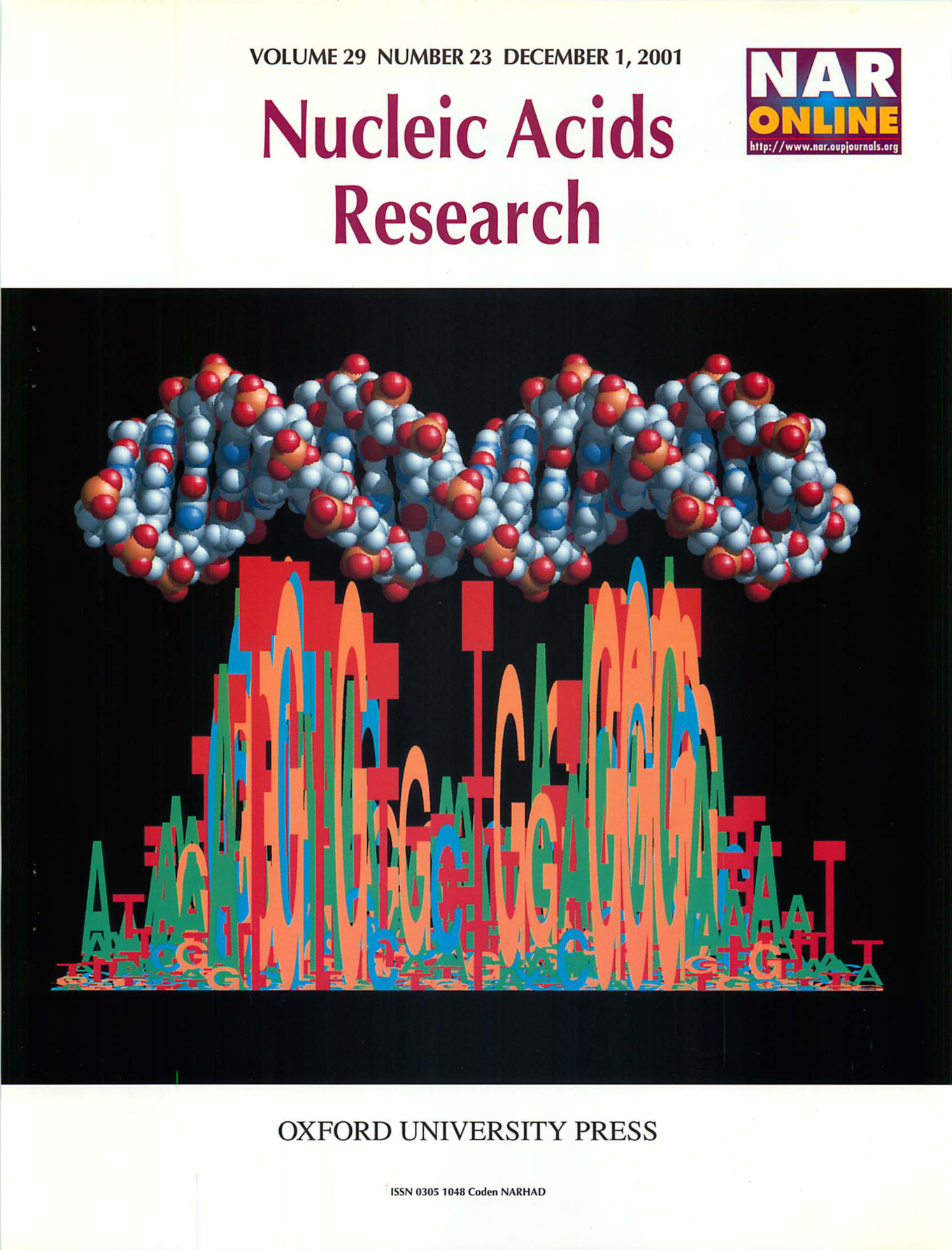
Dubbed "Tom's T" by Dhruba Chattoraj, the unusually conserved thymine at position +7 in bacteriophage P1 plasmid RepA DNA binding sites rises above repressor and acceptor sequence logos. The T appears to represent base flipping prior to helix opening in this DNA replication initation protein. (See papers by Schneider and Lyakhov et al in this issue).

Chen.Schneider-island2006
Z. Chen and T. D. Schneider.
Comparative analysis of tandem T7-like promoter containing regions
in enterobacterial genomes reveals a novel group of genetic islands.
Nucleic Acids Res.
34:1133-1147, 2006.
https://doi.org/10.1093/nar/gkj511
https://alum.mit.edu/www/toms/papers/t7island/
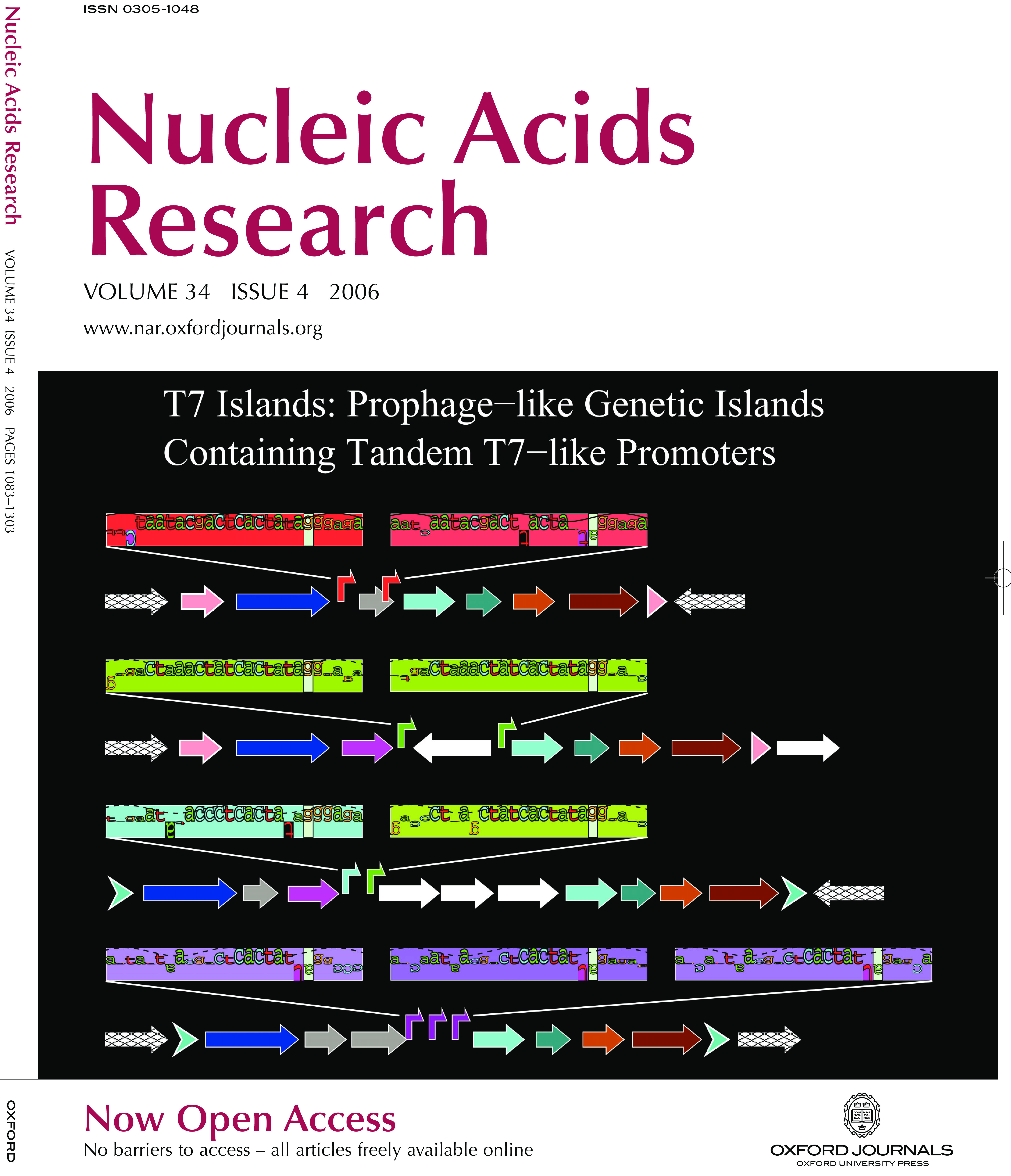
Twelve prophage-like T7 islands have been discovered in pathogenic bacterial genomes. These islands contain two or three tandem T7-like promoters that should be activated when a bacterial cell is infected by bacteriophage T7 or a related phage. The illustration shows genetic maps for four of the islands, Ty2, BS512, E22 and ECA, which are found in the genomes of S. enterica Ty2, S. boydii BS512, E. coli E22 and E. carotovora SCRI1043 respectively. The T7-like promoters are represented by different colored bent arrows (red, T7; green, K1F; cyan, T3; magenta, unknown T7-like) and by corresponding sequence walkers. As in previously known mobile genetic elements, two of the islands, Ty2 and BS512, are adjacent to a tRNA-Gly gene (pink arrows) and have direct repeats of the 3' end of the tRNA gene (pink arrow tips). The other two islands, E22 and ECA, have different direct repeats on their ends (cyan chevron arrows). Each island encodes an integrase (blue arrows), several putative phage-related proteins (other arrows) and often several insertion sequence elements (white arrows).
